胃间质瘤和胃神经鞘瘤的CT鉴别诊断
2018-07-15 佚名 熊猫放射

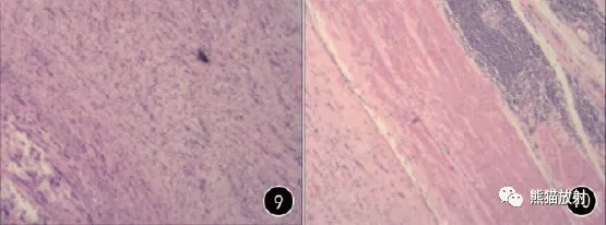
胃窦炎性肌纤维母细胞瘤。男,55岁,黑便3天。a、CT平扫示胃窦部软组织肿块,边界不清,密度不均,中心可见低密度灶,另见小条状钙化灶;b、增强动脉期示病灶边缘明显花环状强化;c、静脉期示对比剂持续填充,呈渐进性不均匀强化;d、未经治疗,第二年病灶的大小、密度、形态均较前无明显变化;e、Ⅱ型梭形细胞密集型(×200,HE)
胃间叶细胞肿瘤约占所有胃肿瘤的3%,主要包括以下四类:
1、平滑肌肿瘤:平滑肌瘤、血管球瘤、平滑肌肉瘤
2、神经源性肿瘤:神经鞘瘤、神经纤维瘤、神经节瘤、副神经节瘤
3、成纤维细胞肿瘤:硬纤维瘤、炎性肌纤维母细胞瘤
4、胃间质瘤
胃肠道间质瘤(gastrointestinal stromal tumor, GIST) 是最常见的间叶源性肿瘤,即便肿瘤很小,也具有潜在恶性,因此,把良性的胃神经鞘瘤(gastric schwannoma, GS) 从GIST中区别开来,对肿瘤治疗方案的选择非常重要。
传统概念上的胃神经源性肿瘤大部分属于间质瘤,现多数学者认为GS 是指免疫组织化学上CD117、CD34、Desmin 和SMA 呈阴性,而S-100呈弥漫强阳性者。而GIST免疫组化CD117 阳性表达。
影像分析的内容主要包括:
肿块大小( 测量肿瘤最大径),发生部位( 贲门、胃底、胃大弯、胃小弯或胃窦),形态( 光滑或有分叶) ,生长方式( 腔内、腔外或混合性生长),囊变( 有或无) ,钙化( 有或无),溃疡( 有或无),强化方式( 均匀或不均匀),渐进性强化( 有或无),肿块周围肿大淋巴结( 有或无),CT值的测量:避开囊变、出血、钙化区。
≤5cm的GIST和GS是最常见也最难鉴别的两种胃间叶源性肿瘤。
GS是一种生长缓慢的神经源性肿瘤,几乎均为良性,恶性者鲜见。
药物伊马替尼( 络氨酸激酶抑制剂) 和手术治疗对GIST均有良好的疗效,但因其具有潜在恶性,术后可能复发和转移,主要的转移部位是肝脏和腹膜后。
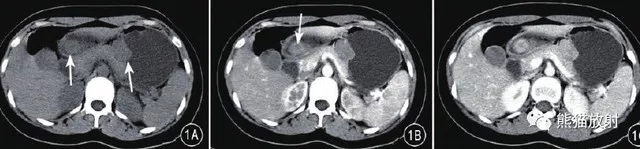
↑ GIST(低危),F24,腹痛、贫血。胃窦及小弯侧各见一软组织肿块,前者不均质强化,后者均匀强化。

↑ GIST(低危),F60,呕血、黑便。胃肿物轻度分叶,可见囊变坏死、钙化,不均质强化,表面溃疡形成。

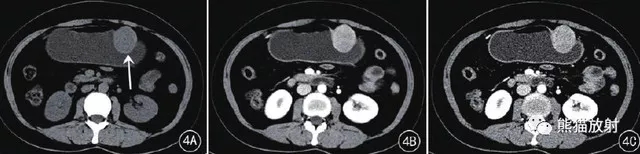
↑ 胃神经鞘瘤,F67。大弯侧卵圆形肿物并溃疡形成,渐进性强化。
↑ 胃神经鞘瘤,F45。明显渐进性强化。
影像表现:
胃体大弯侧为GS的好发部位,GS无分叶;
分叶的出现有利于GIST的诊断。
囊变:
GS一般无囊变,与其他部位的神经鞘瘤不同;瘤内出现囊变时更有利于GIST的诊断,囊变的发生与肿瘤的直径密切相关,直径小于2cm的无囊变。
钙化:
GIST和GS钙化的发生率均较低,GS中出现钙化的概率更小;
溃疡:
两者出现溃疡的概率均较低,GIST相对多见,GIST出现溃疡的倾向于有较高的危险度。
增强扫描:
◆ 静脉期CT值较高、静脉期增强幅度较高,相对有利于GS的诊断;
◆ 肿瘤强化不均时,应考虑GIST;
◆ GS强化均匀,因囊变、出血、钙化而强化不均者少见;
◆ 渐进性强化方式对GS的诊断有提示作用,表现为动脉期病灶强化程度低,静脉期及延迟期多数GS 呈中等或明显延迟强化,实质部分多数为均匀强化。

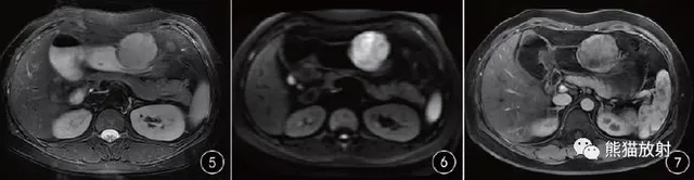
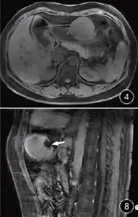
↑ 胃体大弯侧一类圆形软组织密度肿块,同时向腔内外突出,密度较均匀,平扫CT值约29 HU,动脉期病灶轻度强化,门静脉期病灶明显进一步强化,CT值约65HU,较均匀。T1WI 上为等信号图,T2WI 上为均匀稍高信号,病灶与胃黏膜层、浆膜层关系清楚,DWI上病灶为明显高信号,动脉期病灶均匀轻度强化,延迟期呈均匀中度延迟强化,腔内黏膜面可见较大溃疡形成( 白箭) ,溃疡直径约为1.5cm。镜下(HE×200) 见肿瘤组织由梭形细胞组成,局部栅栏状排列,未见明显坏死及核分裂象,周边可见淋巴细胞袖套状聚集( 黑箭) ,肿瘤区域紧贴基底,送检追加切缘阴性。

鉴别诊断:
◆ 胃平滑肌瘤:直径多为3-5 cm,平扫为等密度,密度较均匀,增强扫描轻至中度强化;
◆ 胃癌:肿块表面凹凸不平,与相邻胃组织分界不清,可沿胃壁蔓延,易发生淋巴结及远处转移。


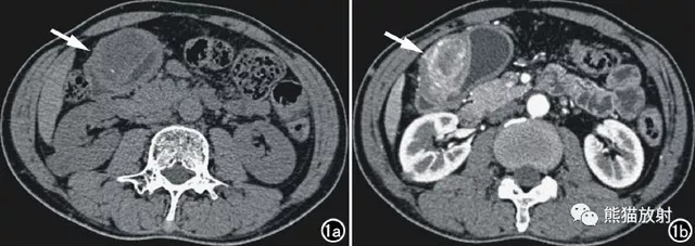
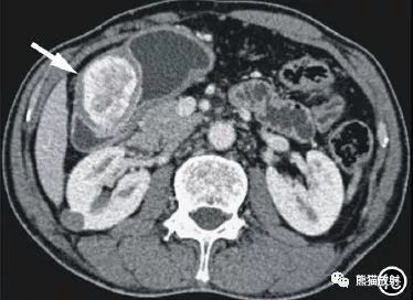
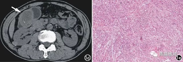
图1 胃窦炎性肌纤维母细胞瘤。男,55岁,黑便3天。a、CT平扫示胃窦部软组织肿块,边界不清,密度不均,中心可见低密度灶,另见小条状钙化灶;b、增强动脉期示病灶边缘明显花环状强化;c、静脉期示对比剂持续填充,呈渐进性不均匀强化;d、未经治疗,第二年病灶的大小、密度、形态均较前无明显变化;e、Ⅱ型梭形细胞密集型(×200,HE)
原始出处:
王健, 徐军良, 厉锋, 胡红杰. CT在(≤5cm)胃间质瘤和胃神经鞘瘤鉴别中的应用价值. 《临床放射学杂志》2017年第02期
小提示:本篇资讯需要登录阅读,点击跳转登录
版权声明:
本网站所有内容来源注明为“梅斯医学”或“MedSci原创”的文字、图片和音视频资料,版权均属于梅斯医学所有。非经授权,任何媒体、网站或个人不得转载,授权转载时须注明来源为“梅斯医学”。其它来源的文章系转载文章,或“梅斯号”自媒体发布的文章,仅系出于传递更多信息之目的,本站仅负责审核内容合规,其内容不代表本站立场,本站不负责内容的准确性和版权。如果存在侵权、或不希望被转载的媒体或个人可与我们联系,我们将立即进行删除处理。
在此留言
本网站所有内容来源注明为“梅斯医学”或“MedSci原创”的文字、图片和音视频资料,版权均属于梅斯医学所有。非经授权,任何媒体、网站或个人不得转载,授权转载时须注明来源为“梅斯医学”。其它来源的文章系转载文章,或“梅斯号”自媒体发布的文章,仅系出于传递更多信息之目的,本站仅负责审核内容合规,其内容不代表本站立场,本站不负责内容的准确性和版权。如果存在侵权、或不希望被转载的媒体或个人可与我们联系,我们将立即进行删除处理。
在此留言


#神经鞘瘤#
70
#间质瘤#
70
#鉴别诊断#
75
#胃间质瘤#
82
不错
94
学习了.谢谢分享.
1