IJPO:HRCT与3DVR CT可良好描述先天性耳道闭锁合并小耳畸形
2012-12-27 Int J Pediatr Otorhinolaryngol 睿医资讯
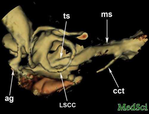
最近一项运用高分辨率CT(HRCT)和三维容积显像(VR)CT探究先天性耳道闭锁合并小耳畸形患者解剖学差异的研究指出,此类患者的面神经,听骨链,鼓室和卵圆窗均有明显异常。利用HRCT和3D VR CT则能很好的显示这些差异,具有重要的临床价值。 &n
最近一项运用高分辨率CT(HRCT)和三维容积显像(VR)CT探究先天性耳道闭锁合并小耳畸形患者解剖学差异的研究指出,此类患者的面神经,听骨链,鼓室和卵圆窗均有明显异常。利用HRCT和3D VR CT则能很好的显示这些差异,具有重要的临床价值。

研究者对43例闭锁患者(34例单侧闭锁,9例双侧闭锁)进行了HRCT检查。而后将HRCT和3D VR检查结果与那些43例病例中PTA结果正常的单侧闭锁患者的正常耳,以及11位伴有感音神经性耳聋但没有相关中耳与内耳发育不全的患者通过独立单样本T检验进行了对比。
结果发现,在HRCT表现上,基线与面神经鼓室段的角度更加锐利。患病耳的听骨链或者锤骨砧骨复合体要小于对照组(P<0.05)。闭锁组的耳道鼓室段更短且鼓室区域更小,同时闭锁组的卵圆窗直径也要小于对照组(P<0.05)。运用3D VR CT成像则可以很好的描述听小骨,鼓膜和乳突段全长的形态学差异。
Objective
To determine the anatomic differences in patients of atresia by using high-resolution computed tomography (HRCT) and 3D volume rendered (VR) CT.
Methods
High-resolution computed tomography (HRCT) was performed in 43 atresia patients including 34 unilateral atresia patients (n=34, 26 males, 8 females, mean age 13.82 years, range 8–19 years) and 9 bilateral atresia patients (6 males, 3 females, mean age 13.2 years, range 9–19 years). HRCT and 3D VR findings were compared with those in 43 normal ears of the unilateral atresia patients with normal PTA results (n=34, 26 males, 8 females, mean age 13.82 years, range 8–19 years) and 11 patients with sensorineural hearing loss but with no associated aplasia of the middle and inner ear (n=22, 7 males and 4 females, range 8–20.8 years, median age of 13.4 years) by using the independent one sample T test.
Results
On the HRCT images, the angle between the basic line and the tympanic segment of the facial nerve is more acute. And the area of the malleus-incus-joint or the malleus-incus-complex in the diseased ears is smaller than that in the control subjects (P<0.05). The tympanic segment is shorter and the area of the tympanic cavity is smaller in the atresia group, while the diameter of the oval window is also smaller in atresia group than that in the control group (P<0.05). The morphologic differences of the small ossicles and the entire length of the tympanic and mastoid segments can be depicted on a single 3D VR CT image.
Conclusions
The facial nerve demonstrates abnormal lateral and anterior displacement in the CAA patients and the area of the Malleus-incus-joint and the tympanic cavity are significantly smaller, and the oval window is much narrower in the control group. HRCT and 3D VR CT provide valuable information about preoperative planning of patients with CAA. Measurements of all the angles and length serve as useful adjunct measurements in determining surgical candidacy.
小提示:本篇资讯需要登录阅读,点击跳转登录
版权声明:
本网站所有内容来源注明为“梅斯医学”或“MedSci原创”的文字、图片和音视频资料,版权均属于梅斯医学所有。非经授权,任何媒体、网站或个人不得转载,授权转载时须注明来源为“梅斯医学”。其它来源的文章系转载文章,或“梅斯号”自媒体发布的文章,仅系出于传递更多信息之目的,本站仅负责审核内容合规,其内容不代表本站立场,本站不负责内容的准确性和版权。如果存在侵权、或不希望被转载的媒体或个人可与我们联系,我们将立即进行删除处理。
在此留言
本网站所有内容来源注明为“梅斯医学”或“MedSci原创”的文字、图片和音视频资料,版权均属于梅斯医学所有。非经授权,任何媒体、网站或个人不得转载,授权转载时须注明来源为“梅斯医学”。其它来源的文章系转载文章,或“梅斯号”自媒体发布的文章,仅系出于传递更多信息之目的,本站仅负责审核内容合规,其内容不代表本站立场,本站不负责内容的准确性和版权。如果存在侵权、或不希望被转载的媒体或个人可与我们联系,我们将立即进行删除处理。
在此留言


#小耳畸形#
112
#先天性耳道闭锁#
92
#耳道闭锁#
76
#3D#
80
#畸形#
80
#先天性#
89